Post By
Gateway Diagnostic Imaging
Date
March 5, 2025
What Does a Chest CT Scan Reveal About Your Health?
A chest CT scan provides your doctor with detailed images of the organs and structures inside your chest. On your chest CT results, your provider can check on the condition of your lungs, bones, and blood supply.
The results from your chest CT can help them better understand your symptoms, so they can make a more accurate treatment plan for your care.
We will show you everything you need to know about getting a chest CT, including how it can help detect conditions related to the lungs, chest injuries, and respiratory conditions, and what to expect during the procedure.
Understanding the purpose of a chest CT scan
A chest CT scan uses advanced imaging technology, to create clear, cross-sectional images of the lungs, blood vessels, bones, and soft tissues in your chest.
This information can help your doctor:
- Understand your symptoms more clearly.
- Gain insights into the condition of your internal organs and tissues.
- Diagnose conditions such as infections, tumors, or blood clots.
- Guide treatment plans for respiratory issues, injuries, and chronic diseases.
What areas of the chest does a chest CT scan focus on?
A chest CT scan provides detailed images of the structures inside your chest.
Including:
- Lungs and airways
- Blood vessels
- Surrounding bones and soft tissues.
- Lymph nodes and parts of the esophagus.
CT imaging is commonly used to check for issues like infections, tumors, blood clots, or injuries.
How does a chest CT help my healthcare provider to diagnose my condition?
A chest CT scan helps your healthcare provider see the internal structures of your chest in great detail, which can identify abnormalities that might not show up on other imaging tests.
The cross-sectional images captured by a CT scan allow your doctor to pinpoint the size, shape, and exact location of issues like tumors, blood clots, or signs of infection. This detailed view helps guide the most accurate diagnosis and best course of treatment for your condition.
In some cases, a chest CT can also guide procedures like biopsies, or monitor your progress during treatment.
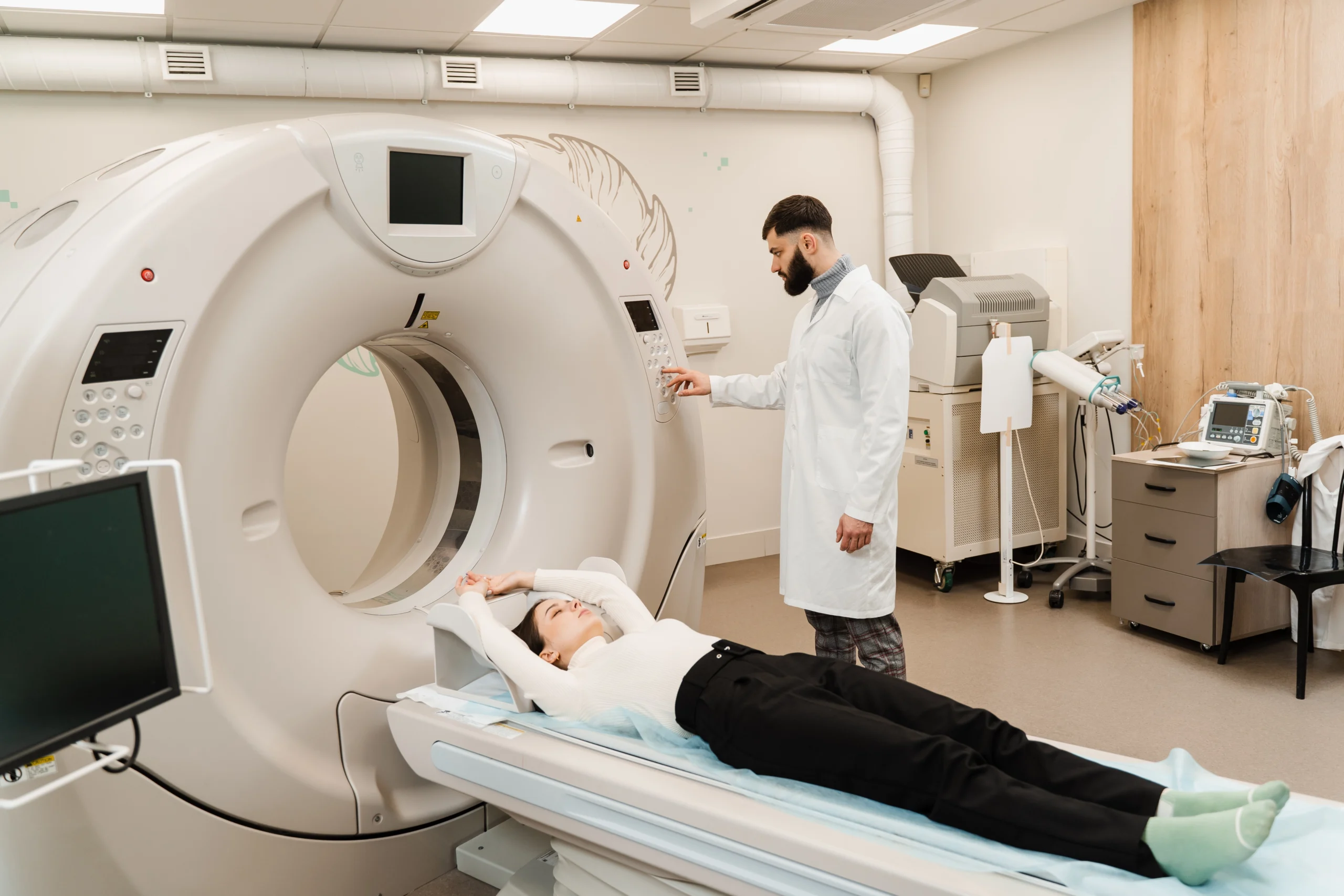
What should I expect during a chest CT scan?
Understanding the process can help ease your mind:
- You’ll lie on a motorized table that slowly moves into a donut-shaped scanner.
- The technologist may ask you to hold your breath briefly to ensure clear images.
- The scan typically takes 30–60 minutes.
- You can communicate with the technologist through an intercom throughout the scan.
Can a chest CT detect lung-related issues?
Yes! One of the benefits of a chest CT scan is that it reveals details of lung-related issues that can be addressed by your healthcare provider.
A chest CT scan can reveal the extent or severity of these conditions and will help your healthcare provider more clearly define a course of action in planning your care.
Chest CT scans are particularly effective at identifying lung-related issues, including:
- Infections: Signs of inflammation, fluid buildup, or dense areas in lung tissue.
- Tumors, growths, or nodules: Detailed images help your doctor locate and evaluate growths.
- Chronic lung diseases: Structural changes caused by conditions like COPD or pulmonary fibrosis.
- Lung consolidation, which appears as dense areas where the air in the lungs has been replaced by fluid or infection.
The scan depicts both the location and extent of tumors, growths, or nodules in the lungs.
How can a chest CT scan identify chronic lung diseases?
A chest CT scan can reveal structural changes in the lungs caused by chronic lung diseases. A trained radiologist can identify structural changes in the lungs that develop over time.
Chest CT scans show scarring, thickened airway walls, and air trapping, which are signs of conditions like chronic obstructive pulmonary disease (COPD) or pulmonary fibrosis. It can also detect small cysts, nodules, and patterns in the lung tissue that are common in some long-term lung conditions.
These detailed images help healthcare providers understand the extent of lung damage and monitor how the disease progresses.
Examining the trauma from a chest injury
A chest CT can provide detailed images of injuries affecting:
- Bones: Detect fractures or displaced bone fragments in the ribs or sternum.
- Soft tissues: Assess damage to muscles, ligaments, or blood vessels.
- Internal organs: Identify punctures, collapsed lungs, or fluid buildup.
Chest injuries can involve not just bones but also soft tissues and vital organs, and a CT scan can provide detailed images to help identify any trauma. Here’s how a chest CT scan can shed light on specific aspects of chest injuries.
How does a chest CT scan help identify fractures or breaks in the ribs or sternum?
The bones of the chest are critical for protecting your lungs and other internal organs, so it’s important to assess them thoroughly after an injury.
A chest CT scan provides highly detailed, cross-sectional images that allow your healthcare provider to pinpoint cracks or breaks that might be difficult to detect otherwise. Beyond locating fractures, a CT scan can reveal the extent of bone displacement, which is when broken pieces of bone move out of alignment.
Also, a chest CT scan can help identify any bone fragments that might have been displaced into surrounding tissues, which could pose additional risks.
This is essential information for determining the best course of treatment, whether that involves rest, immobilization, or further intervention.
What can a chest CT scan reveal about damage to soft tissues such as muscles, ligaments, and blood vessels?
A chest CT scan offers a detailed look at the muscles, ligaments, and blood vessels of your chest, allowing your healthcare provider to assess tears, inflammation, or bleeding that might have occurred.
Any damage to your blood vessels can show up on a CT scan, helping your provider to determine if urgent intervention is needed to restore blood flow or to prevent further complications.
How does a chest CT scan assist in detecting injuries to internal organs?
The lungs are highly susceptible to trauma, and a CT scan can reveal conditions like punctures, a collapsed lung, or fluid buildup. These issues might not always be immediately obvious but can have significant effects if left untreated.
The esophagus, a key part of your digestive system, can also sustain injuries during chest trauma. A CT scan can help detect perforations, swelling, or other signs of damage, ensuring prompt diagnosis and treatment.
Your chest CT results can provide a comprehensive overview of the chest cavity, to detect any internal bleeding or abnormal fluid collections, which might indicate damage to organs.
Can a chest CT diagnose respiratory conditions?
Chest CT scans are invaluable for diagnosing:
- Chronic conditions like asthma, COPD, or emphysema.
- Respiratory infections such as pneumonia or tuberculosis.
- Obstructions in airways caused by mucus, foreign objects, or growths.
These scans help providers determine the best course of treatment for breathing problems.
A chest CT scan can identify opaque areas of the lungs, which may indicate infection, as well as thickening of bronchial walls which may indicate disease of the lungs.
The detailed images can confirm or rule out specific conditions, guiding further diagnostic or therapeutic steps.
What respiratory issues can a chest CT scan help detect?
Conditions like asthma, pneumonia, emphysema, COPD, and tuberculosis can appear as unique abnormalities on a chest CT scan.
It can also detect the presence of growths, infections, or scarring that might interfere with lung function.
A trained radiologist can interpret the scan, and share your CT results with your healthcare provider, who can use them to help shape a course of treatment.
How does a chest CT scan evaluate airway obstructions?
A chest CT scan evaluates airway obstructions by creating detailed images of the airways to identify blockages or narrowing.
It can detect foreign objects, mucus plugs, or growths like tumors that might be restricting airflow. A chest CT also shows any swelling or thickening of the airway walls caused by conditions such as asthma or chronic bronchitis.
The information from chest CT results is especially useful for diagnosing breathing problems that other tests may have difficulty identifying.
What insights can a chest CT scan provide about asthma or COPD?
For asthma, a chest CT scan can reveal airway inflammation, thickened airway walls, or mucus buildup that may be contributing to your breathing difficulties.
For COPD, a CT scan can detect structural changes like damaged air sacs (emphysema), air trapping, or enlarged lung areas that make it hard to exhale fully.
These details help your healthcare provider to tailor your treatments to your condition, so that you can successfully manage it, and achieve the best available outcomes.
How to schedule your scan with us
At Gateway Diagnostic Imaging Centers, we’re dedicated to providing affordable, impeccable quality imaging services. Our specialized radiologists and certified technologists ensure accurate results and a seamless experience.
Reach out to a Gateway imaging center near you, and schedule your appointment today.
